 亲爱的小花帽们,你们还在纠结二维超声引导下神经阻滞技术无法突破么?你还在准备学习或掌握三维引导下神经组织么?而四维超声引导下神经阻滞已经来袭。N. J. Clendenen等制订了一个四维超声引导下神经阻滞的方法。
2D超声因为其快速准确的优势逐步替代传统盲穿神经阻滞技术。然而2D超声下,在超声视窗内我们有时看不到阻滞针的位置以及药物扩散的情况,应运而产生了3D超声引导神经阻滞技术。3D可以立体观察药物的扩散方向和针尖的位置。但是3D超声不能实时的观察药物的扩散情况和针尖的移动情况。4D超声可以完全解决这个问题。然而4D超声引导神经阻滞仍处于研究性阶段,N. J. Clendenen等给予了一个操作标准值得我们学习。通过这个标准化的操作,他们成功的实时观察到阻滞导管放置情况以及药物的扩散情况和药物容积的计算。具体操作如下:
-
Orient the probe in a transverse position to the path of the nerve. Increase the field of view by widening the sector width. Optimize for the best cross-sectional view of the nerve or vessel by adjusting the focus and gain and using XRES and harmonics.
-
Activate x-Plane and “right invert.” The view established in step 1 will appear in the primary window on the left. Using lateral tilt adjust the reference line in the primary window with the track ball so that it intersects the nerve. A longitudinal view of the nerve will be visible in the secondary plane on the right.
-
Insert the needle in line with the center of the probe at approximately a 45- to 60-degree angle. Identify the needle in the primary plane (short axis view) and again use the lateral tilt to bring the needle into view in the secondary plane (long axis view).
-
Advance the needle to the target while keeping the needle in view.
-
Start 4D and select the 4 panel view showing transverse, longitudinal, coronal, and volume view. With the MPR crosshair set to partial, adjust the crosshair so that it lies over the tip of the needle in the transverse, longitudinal, and coronal view.
-
Draw back on the syringe to ensure that the needle is not intravascular and inject a test dose of local anesthetic. Confirm that the local anesthetic spreads appropriately by enveloping the nerve. Inject the total volume if a single dose nerve block is desired or 2/3 of the total local anesthetic dose if inserting a catheter. The spread of the local anesthetic in all four planes (transverse, longitudinal, coronal, and 3D volume) will be seen and the nerve should now be clearly visible due to the fluid-nerve interface.
-
Thread the catheter 2 to 5 cm past the needle tip.
-
Locate the catheter tip by agitating the final 1/3 of local anesthetic and injecting it through the catheter. The agitated local anesthetic will emerge from the catheter tip and appear as a hyper-echoic cloud.
-
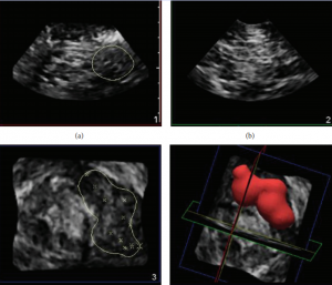
Once the total volume of local anesthetic is given, acquire a data set by pressing “save 3D volume”.
-
Launch Q-lab to measure the approximate volume of local anesthetic in the perineural space.
-
In Q-lab, adjust the threshold to better visualize the fluid/tissue interface. Press “calliper” on the touch screen and outline the local anesthetic spread with the ellipse tool. Use the track ball to adjust the calliper and measure the diameter of the fluid space. The volume calculation appears on the screen.
4D超声引导下神经阻滞作为一个前沿和有挑战性的技术手段,他能实时的跟踪穿刺针的方向和位置,以及观察阻滞导管的植入情况和药物在神经周围的扩散情况及计算目标药物体积。可有效的指导神经阻滞和术后疼痛,减少2D超声的并发症,量化目标神经阻滞的用药量。对局部麻醉的发展有重要意义。
此文作者N. J. Clendenen, 1 C. B. Robards分别来自于美国耶鲁大学麻醉部和梅奥诊所麻醉部。论文发表于Biomed Res Int2014年杂志。
论文资源:http://www.ncbi.nlm.nih.gov/pubmed/24575416
原创文章,如若转载,请注明出处。